
 High End Cardiac Ultrasound Scan Machine")

ME-P30 Best 4D/5D Color Doppler Portable Ultrasound Diagnosis System
Description
ME-P30 Best 4D/5D Color Doppler Portable Ultrasound Diagnosis System
Portable Color Ultrasound Machine
Introducing our cutting-edge ultrasound machine, the ME-P30, equipped with state-of-the-art technology—4D D-Live real skin rendering function. The ME-P30 stands out for its exceptional capability in providing high-resolution 4D imaging. With its advanced features and intuitive interface, medical professionals can accurately assess fetal development, and conduct detailed examinations with unprecedented clarity and precision. Designed to enhance diagnostic accuracy and streamline workflow, the ME-P30 sets a new standard in ultrasound imaging, empowering healthcare providers to deliver superior patient care.
DW-P30 best color doppler portable ultrasound diagnosis system 5D Real skin
The ME-P30 is an intelligent portable 5D ultrasound scan machine designed with the constant goal of providing uncompromising performance at an affordable price. It brings more advances into OB & GYN for the benefit of patients. ME-P30 relies on the realistic Real Skin 5D ultrasound technology and abundant measurement packages to better protect women’s health. ME-P30 will be your best choice if you want more while cost less.
Specialties
- Abdomen
- Vascular
- Cardiology
- OB& GYN
- Urology
- MSK
- Interventional ultrasound
- Small parts
- Anesthesiology
- Pediatrics
- Orthopedics
Real skin rendering
Micron Imaging Technology
Micron imaging technology, real-time tracking of specific signals at the edges of different tissues, to achieve edge enhancement, and monitor each pixel at the same time; optimize the internal signal of the organization and perfectly integrate the edge information and the internal pixel information of the organization to restore the real and delicate, excellent level contrast Two-dimensional image.
Harmonic Imaging Technology(THI)
It improves image clarity by improving tissue contrast resolution, spatial resolution, and eliminating near-field artifacts. It is
mainly used for the diagnosis of cardiovascular and abdominal diseases. It plays an important role in evaluating the lesion area and boundary division of patients with imaging difficulties. The technology has been fully approved by clinicians. Harmonic technology retains the second harmonic signal to
the greatest extent on the basis of removing the fundamental signal, which increases the signal strength by more than 30% compared with the traditional signal processing, reduces noise and artifacts, and improves the contrast resolution of
tissue images.
Trapezoid Imaging
Trapezoid imaging is a kind of expanded imaging, which is transformed into a trapezoid on the basis of the original rectangle, and the left and right sides are expanded to a certain extent, achieving a wider field of view. The principle of ultrasound imaging is to scan the human body with ultrasonic sound beams, and obtain images of internal organs by receiving and processing the reflected signals.
Automatic Spectrum Tracking Measurement Technology
Ultrasound Doppler technology is used in the ultrasound system for examining the heart and arteries and veins. It is necessary to extract relevant parameters from the Doppler spectrogram to evaluate the hemodynamic status of the heart and blood vessels. The disadvantage of manual detection is that the operator’s marking of the peak velocity is
relatively monotonous and time-consuming, with poor repeatability and low estimation accuracy; and during the detection, in order to mark the peak velocity, the operator needs to interrupt the acquisition of Doppler signals, which makes it impossible to estimate in real time. This host contains an automatic envelope detection module, which can automatically track the time-related changes of the peak blood flow velocity and average velocity, and display them in real time on the Doppler spectrogram.
Smooth Workflow
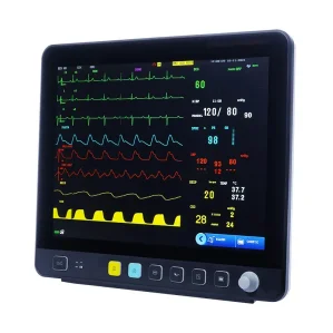
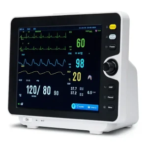
* High resolution medical 15.6 inch display
Brilliant Ergonomics
* High resolution 15.6″LED with tilting functionality
* User-friendly keyboard and controls